

A recent case stopped me.
I uploaded a set of histology images to GPT-5.2 — a dermal lesion with numerous vessels. No clinical history. No immunohistochemistry. Just morphology.
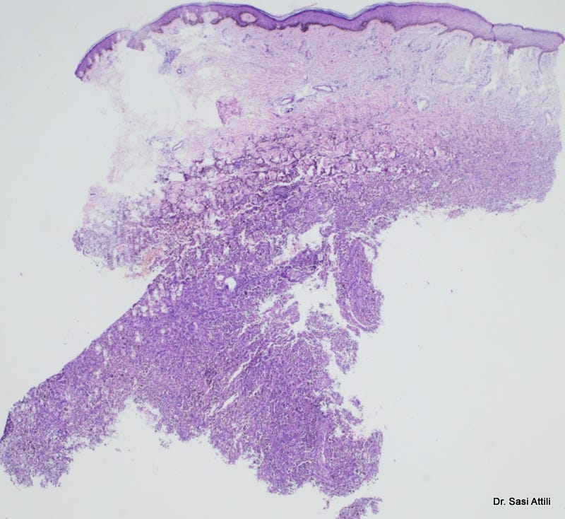
What came back was not a vague answer. It was structured reasoning. Low-power architecture. High-power cytology. A real differential. Systematic exclusion. A final diagnosis: Hemosiderotic dermatofibroma (angiomatoid variant).
It didn’t just label the lesion. It reasoned through it. It noted the absence of cannonball tufts. The absence of endothelial atypia. The fibrohistiocytic stroma. It reframed the lesion as reactive vascular-fibroblastic rather than a primary vascular neoplasm. It even asked the correct follow-up question: Is there peripheral collagen trapping?
That is not trivial pattern matching. That is architectural reasoning.
This is what it concluded:
Brutal conclusion
This lesion behaves morphologically like a fibrohistiocytic tumor with secondary vascular proliferation, not a vascular tumor per se.
If I had to sign it out today:
Hemosiderotic dermatofibroma (angiomatoid variant)
If you have CD34 and FXIIIa, that will settle it in one shot.
If you want, we can go deeper into subtle differentials — but this is not Kaposi territory.
What the AI Did Well
The model separated low-power pattern recognition from high-power assessment — exactly how we are trained. It constructed a meaningful differential and ruled entities out using explicit morphologic criteria. It suggested appropriate immunohistochemistry (CD34, Factor XIIIa). The reasoning was coherent and disciplined.
In purely morphological terms, the performance would not embarrass a registrar.
That matters.
What It Could Not Do
But here is the boundary.
The images were curated. High quality. Representative. I selected them.
AI does not know when the biopsy is inadequate. It cannot detect sampling error, interpret artefact in context, question fixation quality, or request deeper levels because something “feels wrong.” It sees only what it is shown. It does not know what is missing.
That limitation is structural, not temporary.
It also lacks clinicopathological correlation. Age, immunosuppression, medication history, lesion evolution — these are not decorative details. They shape interpretation. Morphology in isolation is powerful, but it is not the whole diagnostic act.
And then there is a subtler risk: automation bias. When a system produces confident, structured reasoning, humans tend to defer to it — even when their own intuition hesitates. The danger is not incompetence. The danger is persuasive competence.
Threat or Tool?
Here is my position.
GPT-5.2 is the most capable AI diagnostic assistant I have encountered in dermatopathology. As a morphological second opinion, it is already useful. It can validate reasoning, surface overlooked differentials, and reduce blind spots.
But diagnosis is not only morphology. It is judgment under uncertainty. It is recognising when the specimen is the problem. It is integrating context. It is taking responsibility.
AI can assist analysis. It cannot assume accountability.
Dermatopathologists will not be replaced by this technology. But dermatopathologists who ignore it may find themselves outpaced by those who understand both its power and its limits.
The question is no longer whether AI belongs in pathology.
The question is whether we engage with it critically — or allow it to shape practice without us.
Dr. Sasi Kiran Attili is a consultant dermatopathologist practising at Visakhapatnam, India. This post reflects personal clinical observations and does not constitute diagnostic guidance.