

Anatomical boundaries, mosaicism and shared immune mechanisms: questioning the biological divide between segmental and nonsegmental vitiligo.
The Article in 30 Seconds
The Hype: A sharply bounded unilateral patch is readily labelled segmental vitiligo (SV). Bilateral disease becomes nonsegmental vitiligo (NSV). SV has anatomical segments and a distinct immune mechanism, while NSV is bilateral and biologically separate.
The Evidence: Our 2016 study identified recurring, sometimes complementary anatomical patterns in NSV. Sarma subsequently found recognizable developmental patterns in 58.6% of his NSV cohort. Two independent studies challenge the assumption that NSV lacks anatomical segmentation, although neither establishes genetic mosaicism.
My Verdict: The labels are clinically useful, but they can encourage a questionable inference: that anatomical organisation belongs chiefly to SV. Anatomical patterning is also observed in NSV.
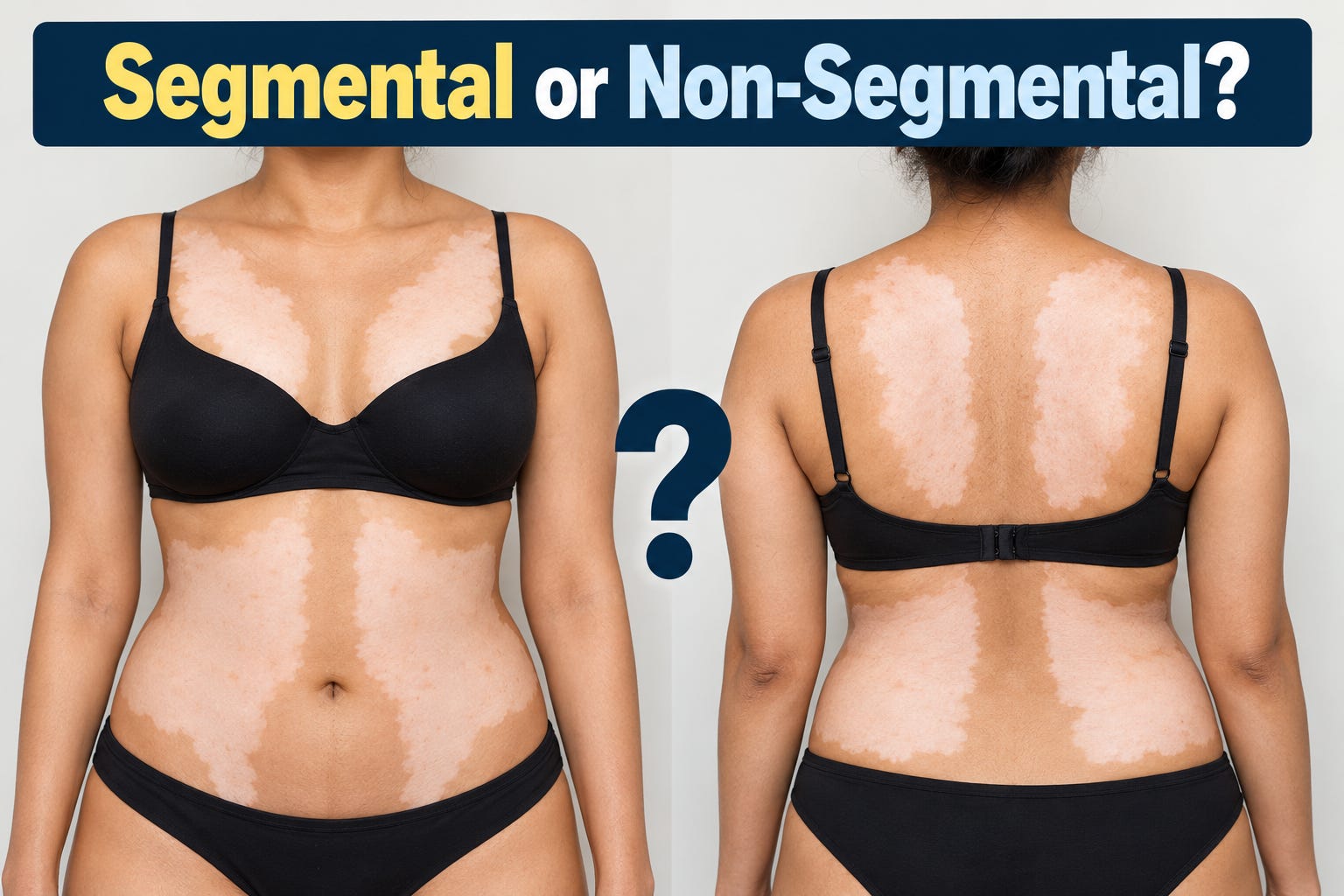
In 2016, we examined more than 3,500 photographs from 615 patients, including 151 with SV and 464 with NSV. We described recurring divisions around the mouth and eyes, at the wrists and across the trunk. Some patients showed apparently complementary patterns: an area depigmented in one was spared in another.
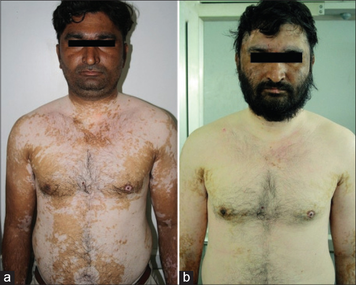
Figure 5 captured something a single photograph cannot: over two years, previously conspicuous truncal boundaries disappeared as vitiligo spread towards near-universal involvement. An evolving lesion may reveal an apparent anatomical territory at one stage and obscure it at another.
What Sarma got right—and overlooked
In 2020, Nilendu Sarma reported patterns resembling Blaschko’s lines or embryonic pigmentary territories in 154 of 263 patients (58.6%) with bilateral NSV involving the face and neck. We disputed the suggestion that patterned NSV had not previously been described. His published reply then criticised our methods.
More importantly, his assertion that we had overlooked Blaschkoid patterns was factually incorrect. Our 2016 paper explicitly described facial patterns consistent with Blaschko’s lines and illustrated evolving Blaschkoid lesions on the trunk in Figure 10. Our observations also extended to broader anatomical divisions and complementary patterns of affected and spared skin.
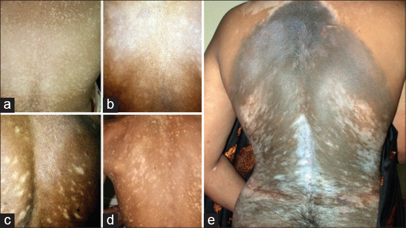
Sarma correctly noted that our study did not quantify the frequency of each proposed segmentation or validate the templates against independent controls. These limitations affect the strength of the conclusions, but they do not negate the observations or their publication priority.
His own study reported recognizable patterns in 58.6% of patients, a useful quantitative addition. However, the anatomical correspondence was also assessed visually, without molecular confirmation of mosaicism.
Both investigations support the possibility of developmentally patterned susceptibility in NSV. Our work documented this phenomenon earlier and across multiple body regions. Sarma subsequently provided additional observations using a more restricted anatomical framework.
Same immune target, different systemic signals?
Our 2013 biopsy series found microscopic inflammation in 78% of 50 SV cases and 70% of 154 generalised cases. Later, Willemsen and colleagues compared blood from 12 patients with stable SV and 22 with stable NSV. Circulating regulatory T cells were reduced in NSV, especially among patients with accompanying autoimmune disease; antibodies directed against tested melanocyte antigens were found in 8/22 NSV patients and none of the 12 SV patients. Consider discoid and systemic lupus: patients with skin-limited discoid lupus may have negative or low-titre ANA despite overlapping immune pathways with systemic lupus. Their manifestations and clinical risks nevertheless differ considerably. Similarly, SV and NSV may share melanocyte-directed cytotoxic mechanisms even when systemic immune markers and disease course differ. Whether their initiating events are also shared remains unresolved.
Anatomical segmentation extends beyond SV
The idea that vitiligo may follow developmentally determined territories is hardly new. Taïeb proposed somatic mosaicism in SV in 2008. Kinsler and Larue subsequently suggested that different embryonic melanocyte populations might explain segmental and bilateral pigmentary patterns. Oh and Oh revisited mosaicism in 2026. These hypotheses differ in their proposed mechanisms, but all recognise the possible importance of developmental anatomy.
Benzekri and colleagues offered another explanation, reporting excellent correspondence with arterial territories in 72% of 140 SV cases. However, the subjective matching, absence of an NSV comparator and lack of chance-controlled analysis limit the conclusions. Vascular anatomy is one possibility among several; developmental, neural and local immune factors may also determine regional susceptibility.
The debate has become overly concerned with identifying the correct mechanism before acknowledging the clinical observations.
Molecular confirmation is not a prerequisite for recognising anatomical patterns.
Future research must determine how reproducible these boundaries are, whether they exceed chance correspondence and what biological mechanisms produce them. Meanwhile, the SV–NSV distinction remains valuable for predicting clinical behaviour and guiding treatment.
The available evidence already challenges the assumption that segmentation belongs exclusively to SV.
Anatomical patterning is also observed in NSV.
What remains unresolved is its biological basis.
References and Further Reading
Attili VR, Attili SK. Anatomical segmentations in all forms of vitiligo: A new dimension to the etiopathogenesis. Indian J Dermatol Venereol Leprol. 2016;82:379–388. https://doi.org/10.4103/0378-6323.181463
Sarma N. Nonsegmental vitiligo follows Blaschko’s lines and embryonic pigmentary segments. Indian J Dermatol Venereol Leprol. 2020;86:350–358. https://doi.org/10.4103/ijdvl.IJDVL_790_18
Attili VR, Attili SK. Comments on: “Nonsegmental vitiligo follows Blaschko’s lines and embryonic pigmentary segments.” Indian J Dermatol Venereol Leprol. 2020;86:683–684. https://doi.org/10.4103/ijdvl.IJDVL_873_20
Sarma N. Author’s reply. Indian J Dermatol Venereol Leprol. 2020;86:685–687. https://doi.org/10.4103/ijdvl.IJDVL_1026_20
Attili VR, Attili SK. Segmental and generalized vitiligo: Both forms demonstrate inflammatory histopathological features and clinical mosaicism. Indian J Dermatol. 2013;58:433–438. https://doi.org/10.4103/0019-5154.119949
Willemsen M, Post NF, van Uden NOP, et al. Immunophenotypic analysis reveals differences in circulating immune cells in the peripheral blood of patients with segmental and nonsegmental vitiligo. J Invest Dermatol. 2022;142:876–883.e3. https://doi.org/10.1016/j.jid.2021.05.022
Garelli CJ, Refat MA, Nanaware PP, et al. Current insights in cutaneous lupus erythematosus immunopathogenesis. Front Immunol. 2020;11:1353. https://doi.org/10.3389/fimmu.2020.01353
Taïeb A, Morice-Picard F, Jouary T, et al. Segmental vitiligo as the possible expression of cutaneous somatic mosaicism: Implications for common non-segmental vitiligo. Pigment Cell Melanoma Res. 2008;21:646–652. https://doi.org/10.1111/j.1755-148X.2008.00511.x
Kinsler VA, Larue L. The patterns of birthmarks suggest a novel population of melanocyte precursors arising around the time of gastrulation. Pigment Cell Melanoma Res. 2018;31:95–109. https://doi.org/10.1111/pcmr.12645
Oh J, Oh SH. Segmental vitiligo and somatic mosaicism: From pathogenesis to therapeutics. Ann Dermatol. 2026;38:159–166. https://doi.org/10.5021/ad.25.250
Benzekri L, Cario-André M, Laamrani FZ, Gauthier Y. Segmental vitiligo distribution follows the underlying arterial blood supply territory: A hypothesis based on anatomo-clinical, pathological and physio-pathological studies. Front Med (Lausanne). 2024;11:1424887. https://doi.org/10.3389/fmed.2024.1424887