

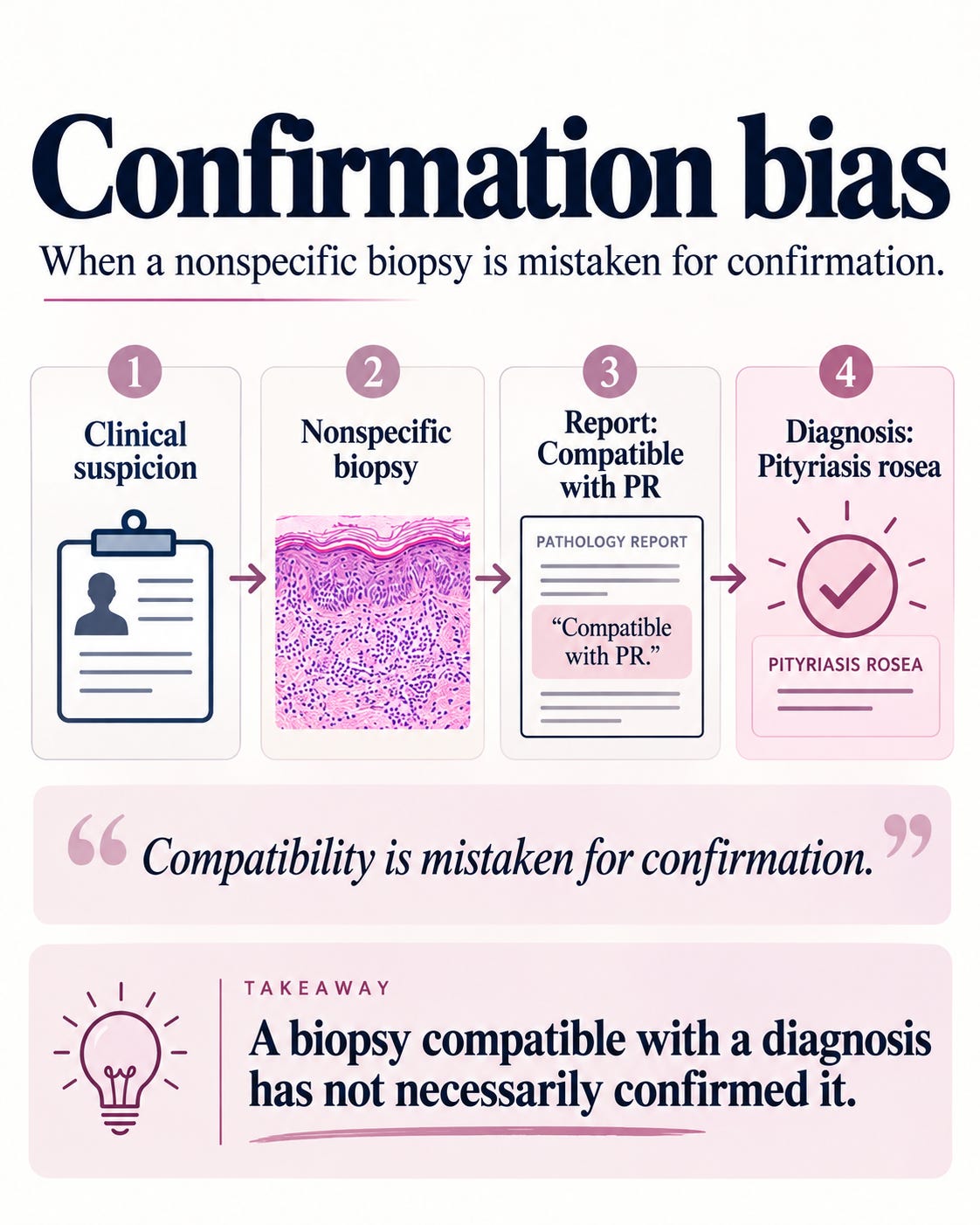
A patient presents with a papulosquamous eruption. The dermatologist suspects pityriasis rosea (PR), but isn’t entirely convinced. A biopsy is requested, presumably to settle the matter.
The dermatopathologist finds mild superficial perivascular lymphocytic inflammation, perhaps some focal spongiosis, and little else.
The report reads: “Nonspecific superficial perivascular dermatitis. Features consistent with pityriasis rosea in the appropriate clinical setting.”
The clinician is reassured. Histopathology has apparently confirmed the diagnosis. How splendid!
Except that the biopsy has confirmed nothing specific…..
It has demonstrated a nonspecific inflammatory reaction that could accompany PR, eczema, a drug eruption or several other dermatoses. It has neither established PR nor necessarily excluded the alternatives.
The clinician suspected pityriasis rosea. The pathologist agreed. The biopsy contributed little. Two opinions, one assumption—and suddenly we have a diagnosis.
The mythology of characteristic histology
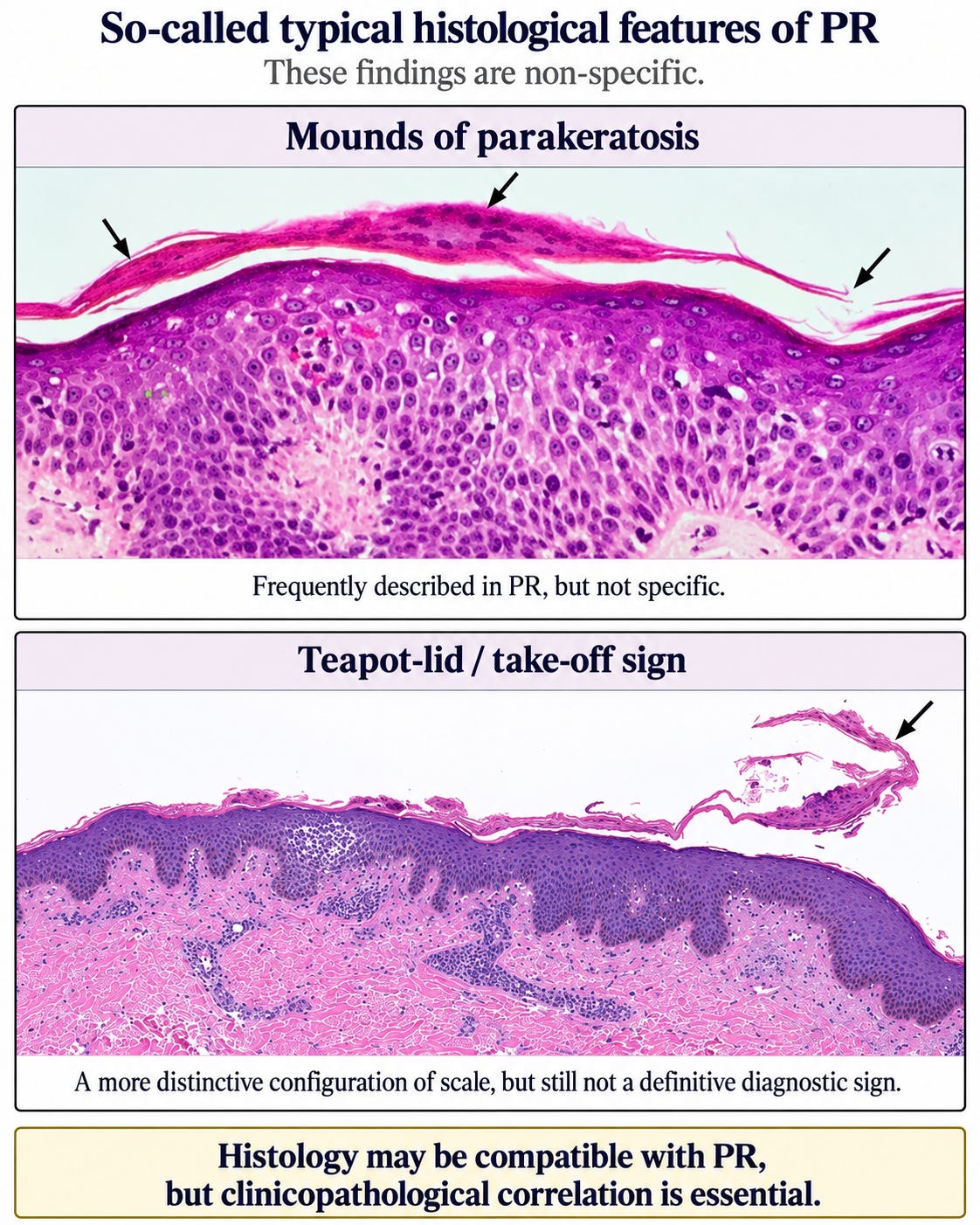
Mounds of parakeratosis: Much ado about nothing?
Mounds of parakeratosis have somehow acquired an exalted position in the histopathology of pityriasis rosea (PR). But why should focal parakeratosis be specific to PR?
Even Thomas and Khopkar, while promoting the salute sign, acknowledged similar mounds in subacute eczema, eruptive psoriasis, pityriasis lichenoides chronica and patch-stage mycosis fungoides. Ernst and Giubellino documented them in maculopapular drug eruptions. Cardoso and colleagues described mound-like parakeratosis in seborrhoeic dermatitis and early psoriasis.
Apparently, parakeratosis has not signed an exclusive contract with pityriasis rosea.
Yet we continue to teach these mounds as though they possess some mysterious diagnostic affinity for PR. Where are the properly validated sensitivity, specificity and likelihood ratios?
A mound is a mound. Giving it a prestigious association does not make it a diagnostic criterion.
I have discussed the related problem of pityriasiform spongiosis elsewhere. Giving a nonspecific reaction pattern a disease-associated name does not make it disease-specific.
The salute, the teapot and the take-off: When scale develops ambition
And then there is the celebrated salute sign, also variously described as the teapot-spout sign, teapot-lid sign and take-off sign.
A little parakeratotic scale lifts away from the epidermis, and suddenly we have kitchenware, military honours and aviation. Dermatopathology has rarely been so adventurous.
Chuh and colleagues identified the take-off sign in six of eight PR biopsies and none of eight controls. Interesting, certainly. But eight retrospectively selected cases, without documented blinding, hardly establish a definitive diagnostic criterion.
More importantly, what makes the scale lift? No PR-specific pathogenic mechanism has been demonstrated. Cardoso and colleagues themselves acknowledge that detachment of the stratum corneum may be artifactual. Mechanical separation during tissue processing or sectioning therefore remains a plausible alternative explanation.
We are also told that this tiny flap represents the clinical collarette of PR. Really? Where are the systematically mapped biopsies demonstrating that the microscopic flap corresponds to the clinically visible collarette?
Resemblance is not anatomical proof. And the possibility that the finding has acquired diagnostic importance through confirmation bias deserves serious consideration.
A feature does not become specific through repeated appearances in textbooks. Specificity requires comparison with what it is supposed to distinguish.
What is the biopsy actually good for?
Why biopsy pityriasis rosea (PR) in the first place? Presumably, to exclude its clinical mimics, not to admire another example of nonspecific spongiosis.
The differentials clinically may include pityriasis versicolor, atypical dermatophytosis (particularly tinea incognito), guttate psoriasis, parapsoriasis and other viral exanthems.
Can a biopsy actually help?
Convincing psoriasiform changes may favour psoriasis. Demonstrating fungal elements can establish dermatophytosis or pityriasis versicolor. A PAS stain may help reveal otherwise inconspicuous fungi, although the numerous spores and short hyphae of pityriasis versicolor are often readily visible on H&E itself.
But did we really need a punch biopsy to discover a fungus that a simple skin scraping might have revealed?
Parapsoriasis and other viral exanthems are a different matter. Their histological findings may overlap substantially with PR. Even early mycosis fungoides cannot reliably be excluded by a single nonspecific biopsy. And if secondary syphilis is suspected, appropriate serology is essential—a bland biopsy is hardly a substitute for VDRL/RPR and treponemal testing!
And if the histology shows nothing beyond mild spongiosis and superficial perivascular inflammation?- The biopsy has neither confirmed PR nor reliably excluded its mimics.
That is the crucial distinction: a biopsy is valuable when it provides positive evidence for an alternative diagnosis—not when it merely fails to contradict the original clinical suspicion.
A more useful report
When the findings are nonspecific, why not say exactly that?
Descriptive Diagnosis (No specific diagnosis possible): Mild superficial perivascular lymphocytic dermatitis.
Comments: The findings are nonspecific and do not independently establish pityriasis rosea. Clinicopathological correlation is required. Important clinical mimics should be investigated as appropriate.
If there are convincing findings favouring another diagnosis, report them. If not, acknowledge the limitation.
There is no disgrace in a nondiagnostic biopsy. There is considerably less justification for dressing it up as confirmation.
Classical PR rarely needs a biopsy. Atypical PR may warrant one, particularly when an alternative diagnosis would change management. But the decision should be driven by a specific diagnostic question, not by the hope that a microscope will somehow resolve every uncertain eruption.
Before ordering a biopsy, ask what you expect it to distinguish. After examining it, ask whether it actually distinguished anything.
If the answer is no, the most useful contribution may be the simple admission that the histology is nonspecific.
A microscope is a powerful diagnostic instrument. It is not a rubber stamp for the clinical impression.
References and further reading
Özyürek GD, Alan S, Çenesizoğlu E. Evaluation of clinico-epidemiological and histopathological features of pityriasis rosea. Postepy Dermatol Alergol. 2014;31:216–221. doi:10.5114/pdia.2014.40641.
Chuh A, Zawar V, Karad G. A case-control study on the take-off sign in lesional skin biopsies of patients with pityriasis rosea. Iran J Pathol. 2016;11:416–417. Full text.
Flamm A, Merelo Alcocer V, Kazlouskaya V, Kwon EJ, Elston D. Histopathologic features distinguishing secondary syphilis from its mimickers. J Am Acad Dermatol. 2020;82:156–160. doi:10.1016/j.jaad.2019.07.011.
Papp JR, Park IU, Fakile Y, et al. CDC laboratory recommendations for syphilis testing, United States, 2024. MMWR Recomm Rep. 2024;73:1–32. doi:10.15585/mmwr.rr7301a1.
Thomas M, Khopkar U. Salute sign: A nonambiguous histopathological sign in pityriasis rosea. Indian Dermatol Online J. 2016;7:543–544. Full text.
Ernst M, Giubellino A. Histopathologic features of maculopapular drug eruption. Dermatopathology. 2022;9:111–121. Full text.
Cardoso JC, Veraitch O, Gianotti R, et al. ‘Hints’ in the horn: Diagnostic clues in the stratum corneum. J Cutan Pathol. 2017;44:256–278. Full text.