

Dermoscopy has an established role in evaluating skin tumours. Trichoscopy has legitimate applications in alopecia.
But dermoscopy of inflammatory dermatoses—grandly rechristened “inflammoscopy”—deserves more scepticism than it usually receives.
Consider the familiar sequence. A dermatologist takes a history, examines the morphology and distribution, and clinically diagnoses psoriasis. The dermatoscope is then applied. Dotted vessels and white scale appear, and these are presented as confirmation.
There is nothing inherently wrong with that. Every diagnostic test is interpreted within a clinical context. But the important question is not whether dermoscopic findings correlate with psoriasis.
It is whether adding dermoscopy to a competent clinical examination improves diagnosis or management enough to justify the enormous vocabulary that has grown around it.
That remains inadequately demonstrated.
Welcome to the Dermoscopic Safari
Inflammoscopy has produced a spectacular lexicon: dermoscopic Auspitz sign, red globular ring, rail-like scaling, leaf-venation, starry-sky Wickham striae, white starburst, vascular polygons, strawberry patterns, white clouds and orange-yellow structureless areas.
One almost expects the next paper to describe the crescent moon over a misty lake sign.
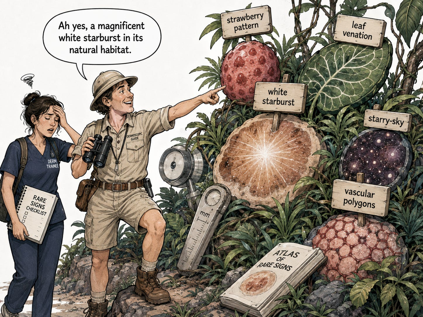
These names are memorable and make excellent conference slides. But metaphors do not create specificity.
“Leaf venation” is still white lines. A “white starburst” represents radial hyperkeratosis and fibrosis. A “strawberry pattern” reflects erythema, follicular plugging and scale.
Much of the inflammoscopic vocabulary therefore describes variations on a relatively small number of biological phenomena: vascular dilatation, scale, crust, follicular plugging, pigment, fibrosis and superficial inflammation. All of these recur across multiple diseases.
Dotted vessels may favour psoriasis; yellow serocrust may favour dermatitis; white network-like structures may favour lichen planus. But none of these observations becomes a diagnosis simply because it has acquired an attractive name.
The major inflammoscopy review itself acknowledges that terminology is variable and metaphorical, that inflammatory disorders often show poorly specific findings, and that dermoscopy should be interpreted after a clinical differential has been established.[1]
That is entirely reasonable.
What remains uncertain is how much the dermatoscope adds once that clinical work has already been done.
Pattern Discrimination Is Not Added Clinical Value
The study by Lallas and colleagues is often cited as strong evidence for inflammoscopy.[2]
It was prospective and methodologically respectable. Dermoscopic readers were blinded to clinical information and histopathological diagnosis, and the combination of regularly distributed dotted vessels, a light-red background and white scale predicted psoriasis with a reported sensitivity of 84.9% and specificity of 88%.
That demonstrates that dermoscopic images contain diagnostically useful information.
But it does not demonstrate that dermoscopy improves the dermatologist’s diagnosis.
The study did not ask clinicians to examine the same patients, record a diagnosis or level of certainty, then add dermoscopy and determine how often the diagnosis became more accurate. Instead, readers were deliberately deprived of the clinical information they would normally have.
They were essentially asked whether dermoscopic features could discriminate among four predefined diagnoses: psoriasis, dermatitis, lichen planus and pityriasis rosea.
That is a valid question. It is simply not the same question as whether dermoscopy adds useful information beyond clinical examination.
A subsequent editorial nevertheless cited this study while stating that dermoscopy had proved significantly superior to clinical examination alone.[3] That comparison was not actually performed.
The claim outran the study design.
And any diagnostic test looks more impressive when it is told that only four diseases are allowed into the room.
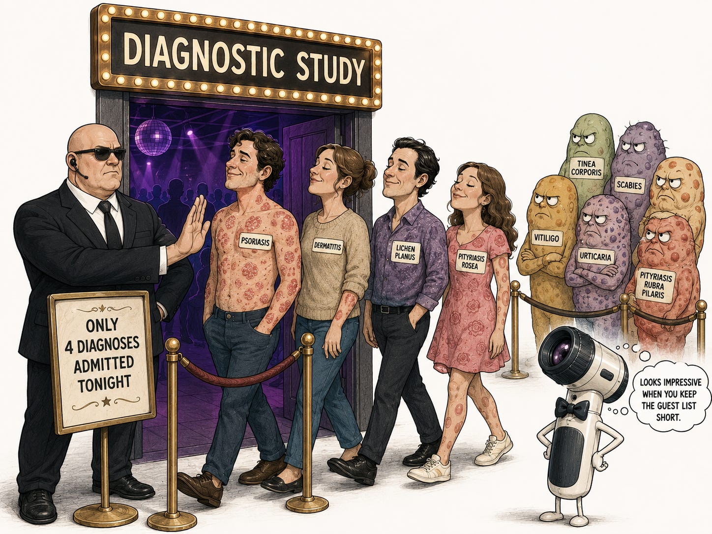
This does not mean inflammoscopy has no role in diagnostically uncertain cases. Within an already restricted differential, regular dotted vessels with diffuse white scale may increase the probability of psoriasis, while patchy vessels and yellow serocrust may push the clinician towards dermatitis.
That is plausible probability modification.
But to establish genuine clinical value, we need studies comparing clinical examination alone with clinical examination plus dermoscopy and asking whether the additional information corrects important diagnostic errors, changes treatment appropriately, reduces investigations safely or improves outcomes.
Much of the existing literature instead establishes associations between dermoscopic appearances and diagnoses that are already known or subsequently confirmed.
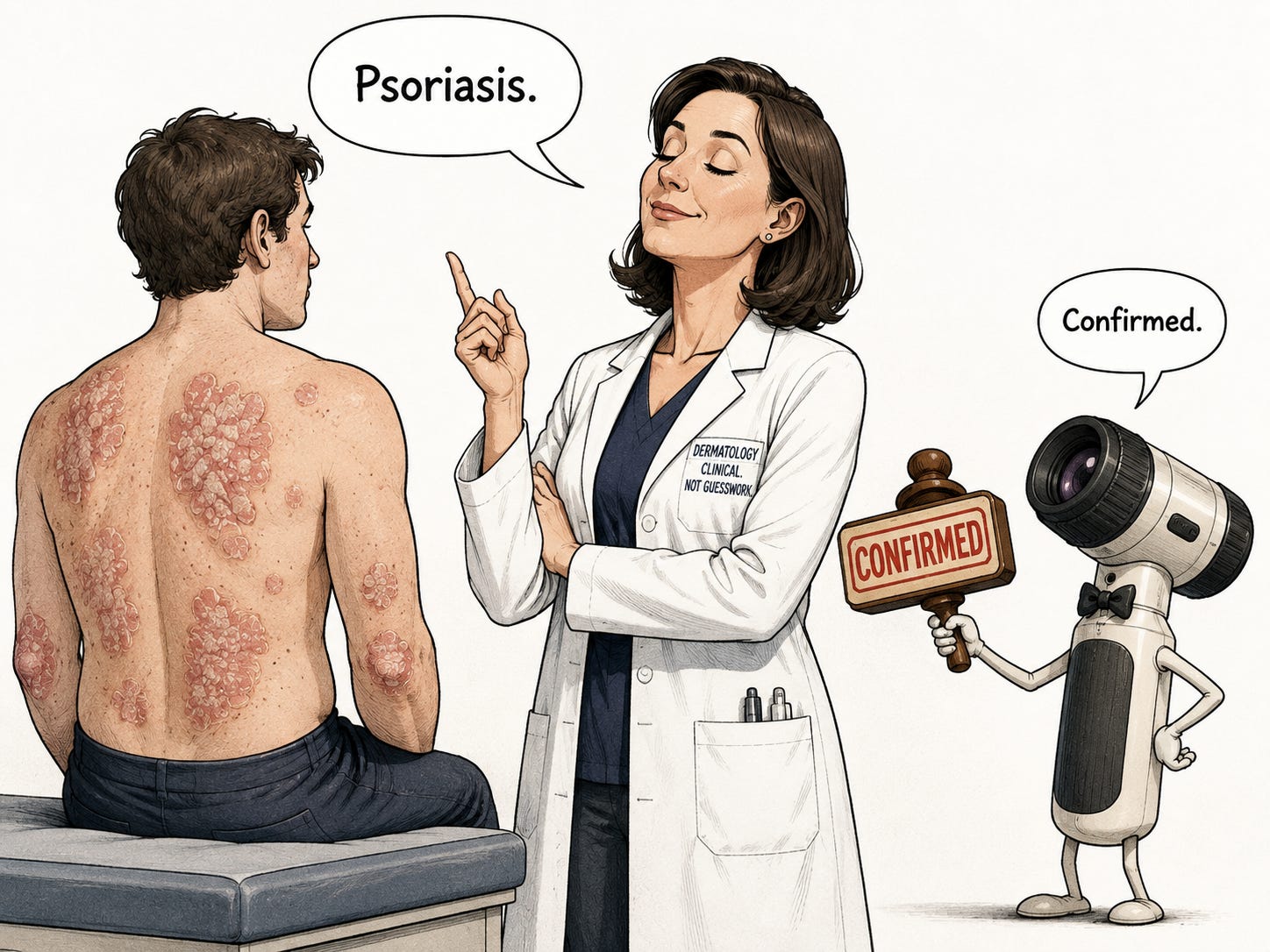
Association is not the same as added clinical utility.
Dermatology Does Not Need More Low-Value Trivia
Dermatology already asks trainees to learn an enormous taxonomy of diseases, variants, synonyms and historical labels while acquiring the skills that actually matter: morphology, distribution, disease evolution, differential diagnosis and clinicopathological correlation.
Inflammoscopy has added another dictionary.
The learner must now remember which diseases supposedly produce strawberries, starbursts, rosettes, polygons, clouds, rail tracks, leaves and starry skies.
For what return?

A systematic review of inflammoscopy in skin of colour identified 85 papers covering 76 dermatoses. Seventy-four were case reports or case series, and only one reached level-III evidence.[4]
That is an extraordinary volume of terminology resting on a remarkably shallow evidence base.
A sign deserves space in the postgraduate brain when it reproducibly improves diagnosis, changes management or prevents harm.
Otherwise, it becomes curricular pollution.
Why the Dictionary Keeps Expanding
This does not require assuming that authors invent signs merely to obtain publications. Their motives cannot be known.
The academic incentive is structural.
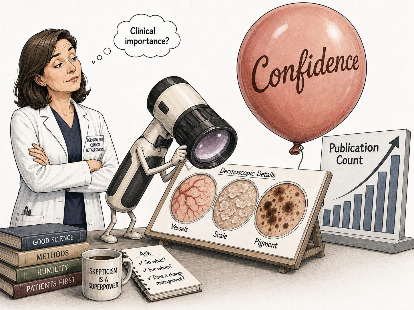
A newly noticed arrangement of vessels, scale and colour can become a case report, correspondence, conference presentation or named sign. External validation is much harder.
A negative study showing that a proposed sign contributes little beyond clinical examination is also less eye-catching than another metaphor accompanied by three attractive photographs.
The predictable result is a literature rich in first descriptions and relatively poor in independent testing.
The system makes it easier to name signs than to prove that they matter.
Did the Dermatoscope Avoid the Biopsy—or Did the Biopsy Validate the Dermatoscope?
Claims that inflammoscopy can reduce “unnecessary biopsies” deserve particular scrutiny.
Pakornphadungsit and colleagues studied 102 patients with inflammatory and infectious dermatoses. Dermoscopic images were interpreted by a blinded dermatologist, and every participant underwent biopsy to establish the diagnosis.[5]
The study did not record whether clinicians intended to biopsy before dermoscopy, whether dermoscopy changed that decision, how many biopsies were actually avoided, whether diagnoses were missed or delayed, or what happened to patients subsequently.
Nevertheless, the authors suggested that dermoscopy might minimise unnecessary invasive investigations.
It might. But that was a hypothesis generated by the study, not an outcome demonstrated by it.
When everyone in a diagnostic study undergoes biopsy, the study can tell us whether dermoscopic findings correlate with histopathological diagnoses.
It cannot tell us whether omitting the biopsy would have been safe.
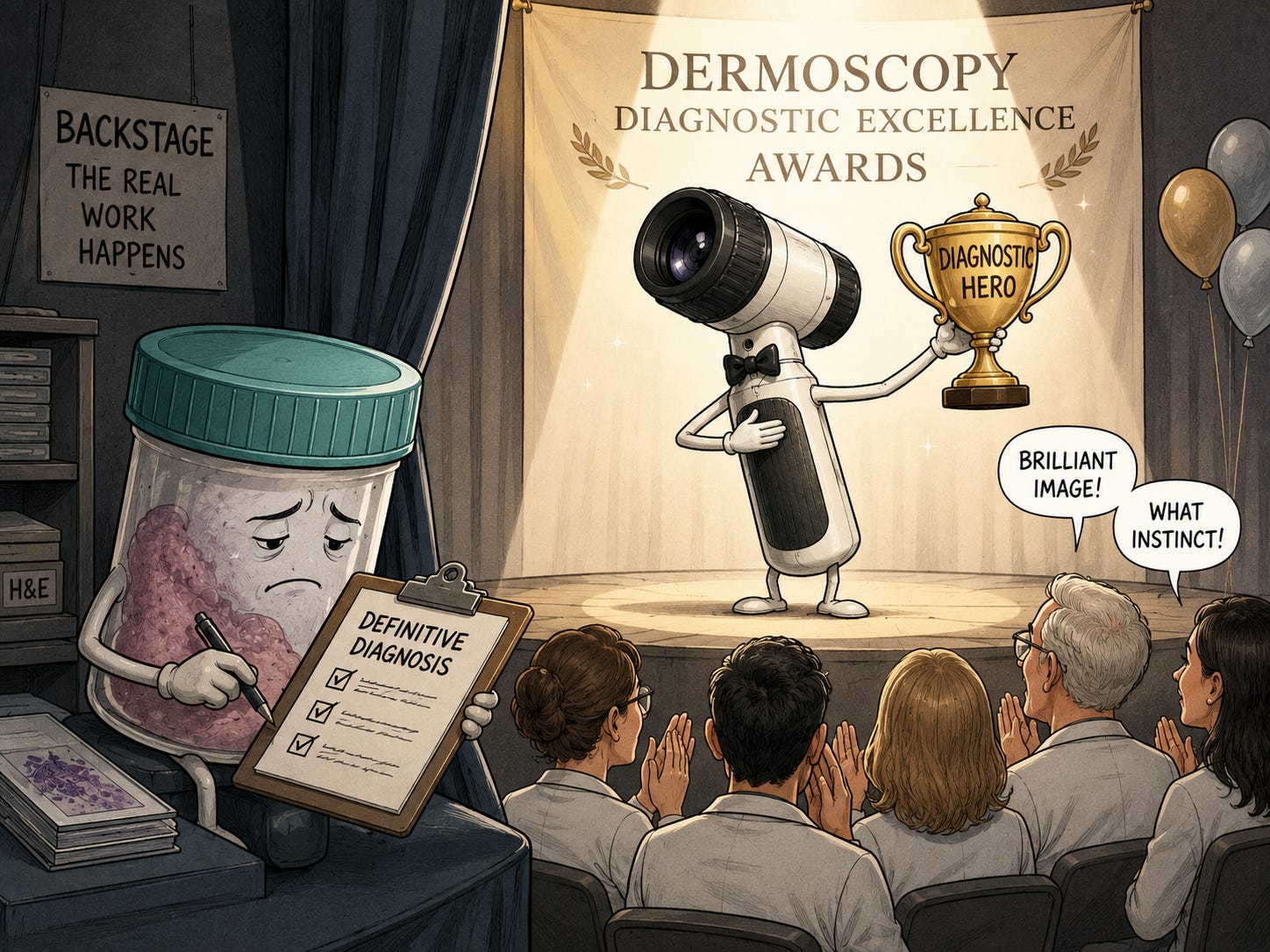
The biopsy did the validating.
Dermoscopy received the applause.
So Where Does That Leave Inflammoscopy?
Inflammoscopy is not invalid because it is interpreted after clinical examination. That is how diagnostic medicine works.
Its problem is that a relatively limited collection of overlapping surface reactions has accumulated an elaborate vocabulary without equivalent evidence that learning this vocabulary materially improves clinical care.
Some patterns probably do modify probability within a restricted differential. That is useful.
But pattern association is not incremental benefit. Greater diagnostic confidence is not necessarily greater diagnostic accuracy. And a biopsy not performed is not automatically a biopsy safely avoided.
When the diagnosis is clinically obvious, inflammoscopy may add little beyond terminology. When the diagnosis is genuinely uncertain and the answer will change treatment, prognosis or surveillance, the dermatoscope does not make dermatopathology redundant.
Inflammoscopy certainly magnifies vessels, scale and pigment.
The unresolved question is whether we have also magnified its clinical importance.
References
Errichetti E. Dermoscopy of inflammatory dermatoses (inflammoscopy): an up-to-date overview. Dermatol Pract Concept. 2019;9:169–180. doi:10.5826/dpc.0903a01.
Lallas A, Kyrgidis A, Tzellos TG, et al. Accuracy of dermoscopic criteria for the diagnosis of psoriasis, dermatitis, lichen planus and pityriasis rosea. Br J Dermatol. 2012;166:1198–1205. doi:10.1111/j.1365-2133.2012.10868.x.
Lallas A, Argenziano G. Dermatoscope—the dermatologist’s stethoscope. Indian J Dermatol Venereol Leprol. 2014;80:493–494. doi:10.4103/0378-6323.144141.
Sławińska M, Żółkiewicz J, Behera B, et al. Dermoscopy of inflammatory dermatoses in skin of color: a systematic review by the International Dermoscopy Society “Imaging in Skin of Color” Task Force. Dermatol Pract Concept. 2023;13:e2023297S. doi:10.5826/dpc.1304S1a297S.
Pakornphadungsit K, Suchonwanit P, Thadanipon K, et al. Dermoscopic features and their diagnostic values among common inflammatory and infectious dermatoses: a cross-sectional study. Clin Cosmet Investig Dermatol. 2023;16:211–220. doi:10.2147/CCID.S397212.